The human eye is a limited organ. The portion of the electromagnetic spectrum that we can see is about 0.0035 percent of the total light in the universe. Without any aid, a normal human eye with 20/20 vision can clearly view up to only about five kilometers (about three miles) in the distance and can distinguish an object as small as about 0.1 millimeter. Just as spyglasses and telescopes extended our range of sight across Earth and into the cosmos, light microscopes allow us to peer at scales hundreds of times smaller than we would otherwise be able to detect. Such technology has bred innumerable discoveries in medicine, biology, geology and plant science.
For 46 years, camera company Nikon has run its Small World contest, which prizes excellence in photography at the tiniest scales—achieved with the aid of the light microscope. Scientists make up a substantial proportion of contest entrants because their work naturally lends itself to stunning visualization. Below is this year’s first-place winner and our editors’ picks for the best images. As entrant Jason Kirk of the Baylor College of Medicine says, the contest is a “unique opportunity to celebrate the convergence of art and science. Images like the ones showcased here are a [wonderful] bridge between the scientific community and the general public.”
This year’s winning image of a juvenile zebra fish was captured as part of research by a team at the National Institutes of Health. They discovered that zebra fish have lymphatic vessels inside their skull—a feature previously thought to only occur in mammals. Such a discovery could expedite and revolutionize research related to neurological diseases such as Alzheimer’s. The researchers stitched together more than 350 individual images to create this single one.
Radula, or “tongue,” of a freshwater snail, stained and captured as a stack of images. The realm of tiny animals is replete with bizarre and sometimes alien forms, says the image’s creator, Igor Siwanowicz of Howard Hughes Medical Institute, who obtained the snail from his lab mate’s aquarium. “It’s a snail’s tongue, looking like a decadent rococo chandelier,” he adds. The image won third place.
Scale from the wing of a blue emperor butterfly (Papilio ulysses). Photographer Yousef Al Habshi, says the challenge in creating this image was finding the correct focal balance between the camera and the scales to capture the light, avoiding overexposure or underexposure.
Daphnia, a water microorganism. To create this image, photographer Paweł Błachowicz used the reflected-light technique: light bounces off the subject and is captured by the camera. This method is usually reserved for opaque objects, Błachowicz says, so he was surprised at this striking outcome. “Daphnia is a transparent organism, and despite this, with the reflected-light technique, it looks astonishing,” he adds.
Crystals formed after heating an ethanol-and-water solution containing L-glutamine and beta-alanine. The proportions of both amino acids must be precisely balanced in order to form such striking crystal structures, says photographer Justin Zoll. He used a polarized-light filter to capture this image, which won 13th place.
Lateral view of a leaf-roller weevil (Byctiscus betulae). The hard exoskeleton is highly reflective and therefore challenging to capture, says photographer Özgür Kerem Bulur, who had to balance the light properly in order to capture these rainbow colors. This image won 14th place.
Head of a tapeworm (Taenia pisiformis) from the gut of a rabbit. The angle of this photograph shows the “teeth” on the edge of the parasite’s head that help it embed itself in its host’s digestive tract. “I love the image’s geometrical beauty, its sculptural qualities and its ambiguity,” says image creator David Maitland. “Is it a fossil or something embedded in sandstone? What is it?”
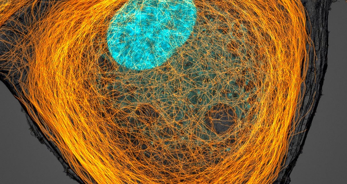
Microtubules (orange) inside a bovine pulmonary artery endothelial cell. The nucleus is shown in cyan. In his work, Jason Kirk of the Baylor College of Medicine uses such cells to benchmark the performance of his microscopy equipment. But the end result, which won seventh place, deserves acknowledgement for its artistic value.
Science in Images is a new category of articles featuring photographs and videos from all the disciplines of science. Click on the button below to see the full collection.


