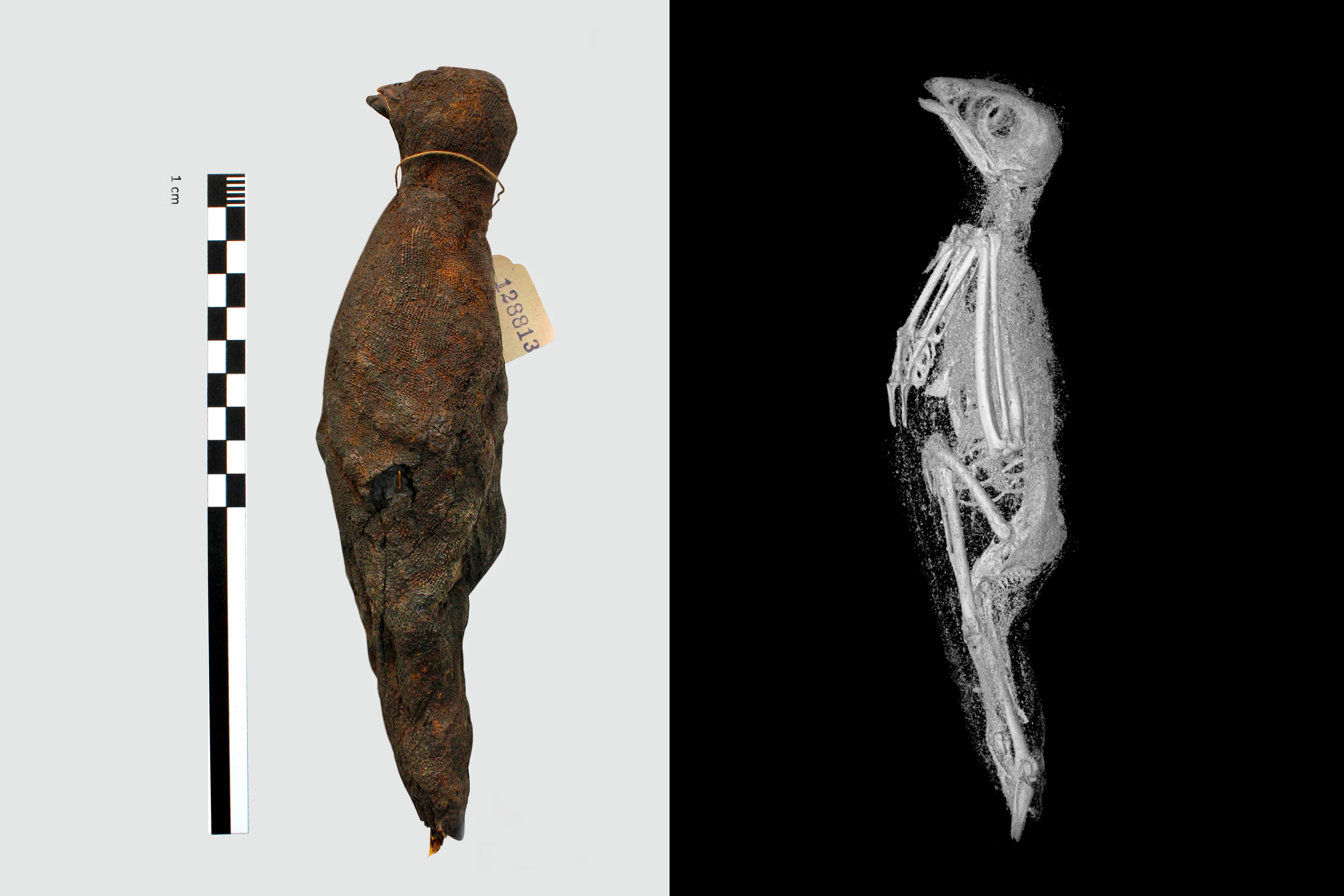
More than 2,000 years ago in Egypt, animals were often mummified as offerings to gods or buried along with human mummies. But many details about the practice are unclear, in part because studying the creatures without damaging them is difficult. Recent Micro CT scans of three animal mummies at Swansea University in Wales offered a more detailed view than ever before. The three-dimensional x-ray images—with 100 times the resolution of a regular medical CT scan—allowed researchers to precisely measure the lengths of the animals’ bones and other features. They reported their findings in August 2020 in the journal Scientific Reports. A cat mummy, for instance, turned out to be a kitten less than five months old, which might have been strangled to death. Measurements of bones in a bird mummy suggest the animal was probably a Eurasian kestrel. And a snake, identified via the scans as a juvenile Egyptian cobra, seems to have been killed by a whipping motion, and its kidneys showed signs that it had been deprived of water during its life. The images also revealed that the snake’s jaw was wide open, and there was trauma in its teeth and jaw bones, suggesting the mouth might have been pried apart during the mummification process. This is the first time such a complex ritualistic process has been documented in a snake, the researchers say.